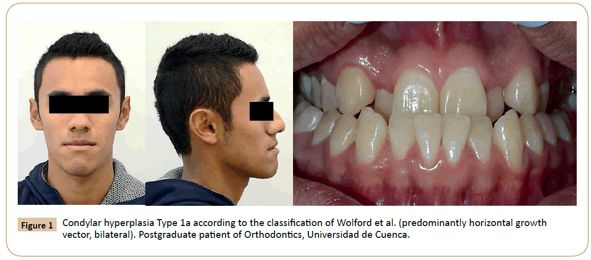
If you have been diagnosed with unilateral condylar hyperplasio, you are probably wondering how to deal with it. Luckily, there are several resources available for you to help you deal with this condition. In this article, we’ll go over the causes, symptoms, and treatment options of unilateral condylar hyperplasia. Read on to learn more!…and be sure to let us know if you have ever experienced any of these conditions!
Case of unilateral condylar hyperplasia
A case of unilateral condylar hyperpasia has been reported in the literature. It is a self-limiting, progressive deformity of the mandible that can affect facial symmetry. The condition is characterized by hyperactivity of the condylar ridges and is classified into two distinct phases – the active phase and the stationary phase. The latter phase is associated with mandibular asymmetry. In addition, skeletal scintigraphy is used to detect this condition, as routine X-rays and CT scans may only detect anatomical changes.
A rare case of unilateral condylar hyperplasis is presented. A 15-year-old female was diagnosed with CH and underwent condylectomy in the right condyle. The active hyperplasia was stopped by surgical reshaping of the condyle. This patient also underwent orthognathic jaw impaction and asymmetric mandibular setback.
The asymmetry in the lower part of the face was found on both CT scans and MR images. A diagnosis of CH was made based on the patient’s history, and the doctor recommended surgical correction. However, the patient refused surgery. Further, the case was not followed up for a long time. Its long-term effects are unknown. If the patient does suffer from CH, treatment should be customized to the individual patient’s unique needs.
Anatomical changes are the most important determinant of the severity of the disease. In this rare case, the condition can be diagnosed in early stages, and early recognition can prevent complications related to abnormal growth. Computer tomography can be used to visualize the structure but lacks functional information. Single-photon emission computed tomography, in contrast, is an excellent tool for detecting active growth changes.
The diagnosis of condylar hyperplasia requires a careful assessment of the patient’s condylar growth state to plan the best treatment for each individual patient. If the patient has an active mandibular condyle, a high partial condylectomy can be performed to correct the facial deformity. Inactive condyles are treated by osteotomy of the upper and lower jaws.
In this case, the condylar neck and head of the mandible were symmetrically enlarged on the left side and the neck was elongated. The 29-year-old mother, who consented to the examination, had facial asymmetry. The medical history of the patient was noncontributory. Clinical examination revealed a deviation of the lower face to the left side and asymmetrical right-side chin. In addition, the prominence of the chin appeared shifted to the left side.
Physiologic bone remodeling of the left condylar head was noted 4 months following the fracture. However, there was no significant improvement of the right condylar hyperplasia. The patient chose to undergo the first treatment option, which was a surgical risk. The patient underwent a proportional condylectomy that involved a 14-mm resection of the left condylar head and rounded the remaining part with a low speed bur. However, the resected condylar head underwent a complete histopathologic evaluation.
Symptoms
One of the main features of unilateral condylar hyperplasia is the progressive enlargement of the mandibular condyle. This disorder affects the mandible, resulting in skeletal asymmetries and psychosocial problems. This disorder is typically unilateral and is the result of trauma or functional influence. Patients with CH should seek surgical reshaping of the condyle to correct the symptoms.
The affected side usually has irregular and thick trabeculae. Trabeculae on the affected side have higher percentages of osteoid compared to the other side. In addition, the affected condyle has an undifferentiated mesenchymal layer and hyperplastic cartilage islands. Patients may have fibro-cartilage and have a hyaline appearance.
Unilateral condylar hyperplasia is a rare disease, with no known cure. There are many causes, including deformities of the mandible and facial bones. Treatment can include surgical removal of the affected joint or appropriate surgical correction. In rare cases, surgery will be needed. A combination of CH and degeneration of the temporomandibular joint may be necessary.
Imaging studies are necessary to confirm the diagnosis of this disease. Computerized tomography and magnetic resonance imaging (MRI) are common diagnostic methods. These imaging studies allow doctors to see the condyle’s asymmetry and approximate when CH started. Imaging studies with radioisotopes are particularly useful for diagnosing condylar hyperplasia because they can identify areas of increased osteoblastic activity.
OPG and CT scan show gross enlargement of the left condyle. MRIs show a slanted mandible, a slanted occlusal plane, and deviation of the dental midlines. The hyperplastic mass of the condyle extends into the lateral preauricular region. The patient can undergo orthodontic treatment if the condition is diagnosed early enough.
Other symptoms of UCH include hemimandibular elongation and hemifacial hyperplasia. Patients may exhibit an open bite. They may also experience facial asymmetry. Patients may also experience pain around the jaw, and they may even experience a clicking sound while opening and closing their jaw. These symptoms may be the result of a benign tumor or malformation of the condyle.
Asymmetric facial morphology is an important diagnostic factor for UCH. It can lead to many facial problems including mandibular asymmetry, jaw deformity, and malocclusion. Single photon emission computed tomography (SPECT) can help diagnose this disease, but it has low sensitivity and is not suitable for routine assessment of growth in CH patients.
Treatment of CH depends on the symptoms of the disorder. A condylectomy may be necessary in severe cases. This treatment, however, can disrupt the TMJ and result in a failure of realignment. An early diagnosis and surgical intervention can improve the patient’s quality of life. If left untreated, condylar resection may result in a permanent defect in the jaw.
Treatment options
While there is no single definitive treatment for CH, various approaches are being used. The gold standard for diagnosis is a clinical examination, followed by radiographic examinations, including bone scintigraphy and single-photon emission computed tomography. Surgical treatment is another option, with the main goal of restoring normal temporomandibular function. Surgical correction has been found to be effective, with minimal effects on the temporomandibular joint.
The etiology of this disorder is unknown, although several theories have been made. It is believed to occur as a result of trauma to the region, triggering a surge in the repair mechanisms and expression of bone forming molecules. Moreover, females are more susceptible to UCH than males, and the condition occurs more frequently in women than men. The good news is that treatment options for unilateral condylar hyperplasia are now more widespread, making them more accessible.
The diagnosis of unilateral condylar hyperplasiy can be confirmed through SPECT scans. 99Tcm-MDP imaging, which was recently used to diagnose 105 cases of unilateral condylar hyperplasia, was performed in 49 patients as a control group. In all affected condyles, 99Tcm-MDP uptake was significantly greater than that of the contralateral side. Further, condyle/skull ratio was higher in the affected side than the contralateral one, which indicates that the underlying bone is active and hyperplastic.
Unilateral condylar hyperplasia is usually diagnosed by SPECT bone scan, and treatment is based on the severity of the deformity, age, and condylar growth activity. Some patients may be treated with unilateral ramus osteotomy of the affected condyle, while others may need bimaxillary surgery. Once treatment options have been determined, the underlying cause must be identified and the best treatment method should be selected.
Diagnostic imaging with SPECT images is the best way to confirm the diagnosis. Using the technetium 99m scan, a single-photon emission computer tomography image of the affected side is performed. An electromyography (EMG) scan of the face can also confirm the diagnosis of unilateral condylar hyperplasia. Using Technetium 99 m-methyl diphosphonate in imaging, the doctor can accurately detect the condylar growth centers.
Orthognathic surgery is another option. This surgery involves removing the affected condyle, which prevents the condition from relapsing. However, it is crucial to note that this is not a permanent cure for unilateral condylar hyperplasia. In some cases, a patient will still experience asymmetric facial appearance. However, a patient’s facial appearance can be restored.
While a number of diagnostic tests are available for CH, SPECT is the most accurate. In fact, a retrospective study of UCH patients revealed that this method had a cut-off value of 55%. Interestingly, a study of patients with UCH revealed that the presence of a hypertrophic layer in the chondrium was correlated with an active form of CH.


